Doktor Made ermittelt

Quelle: KURIER.at, 8. November 2024

Von Birgit Seiser

Nachgefragt. Immer noch ist offen, wann der Doppelmörder Roland Drexler gestorben ist. Experte Mark Benecke erklärt, welche Faktoren für die Bestimmung des Todeszeitpunkts essenziell sind.

Fünf Tage lang suchte eine Hundertschaft an Polizisten Wälder im oberösterreichischen Bezirk Rohrbach (OÖ) nach dem mutmaßlichen Doppelmörder Roland Drexler ab. Am Samstag stand fest, dass er tot ist, seine Leiche wurde in seinem Jagdgebiet entdeckt. Mit Spannung erwartete das ganze Land das Ergebnis der Obduktion, das aber weiter Fragen offen ließ. Der genaue Todeszeitpunkt konnte nicht bestimmt werden. Nun sollen forensische Entomologen das Rätsel lösen.

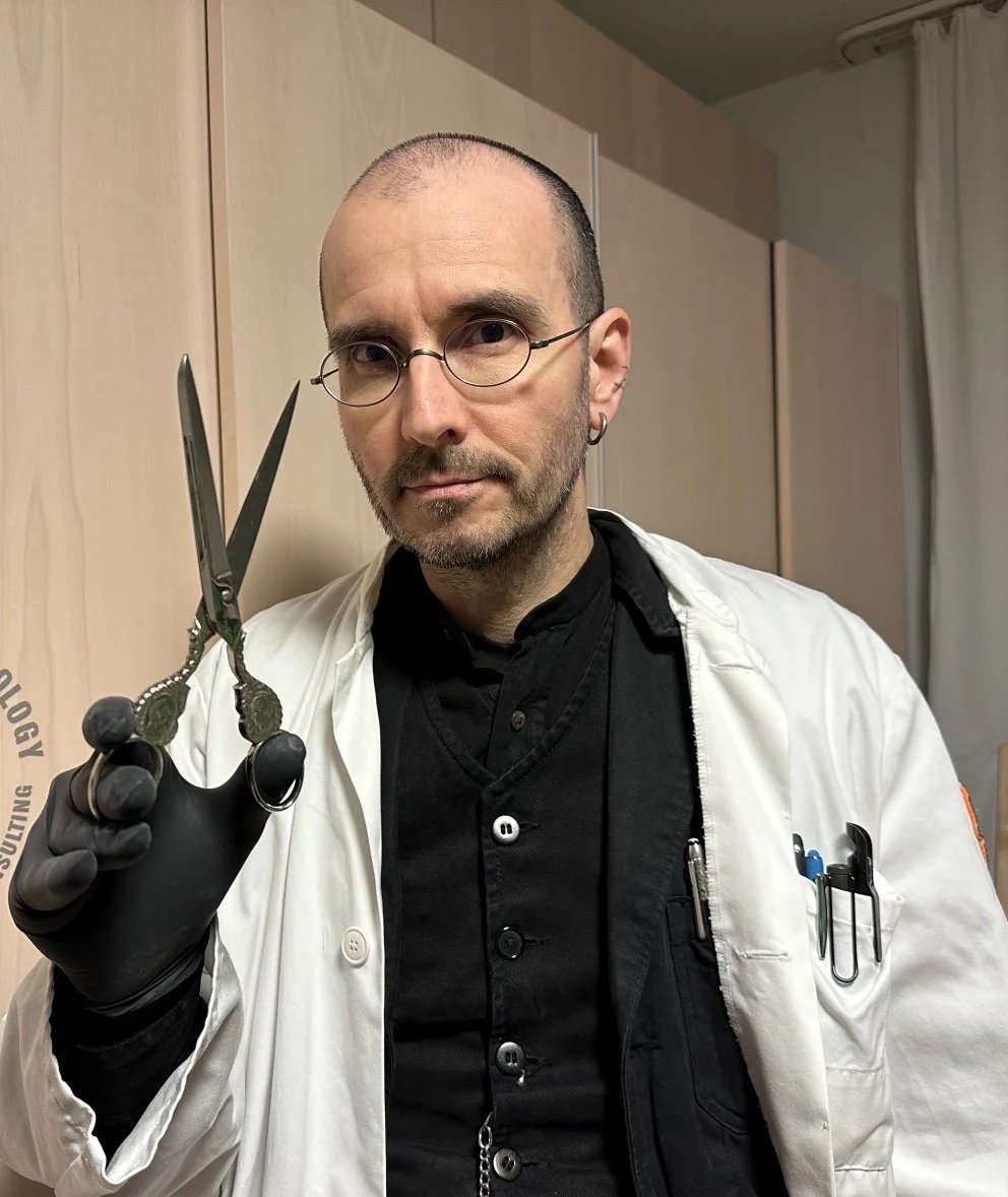
Einer der bekanntesten Vertreter dieses Fachs ist Dr. Mark Benecke. Der Kriminalbiologe, der auch als "Dr. Made" bekannt ist, erklärt im KURIER, wie man den Todeszeitpunkt bestimmen kann und wie Insekten dabei helfen. Die folgenden Zeilen sind nichts für Menschen mit schwachen Mägen.

Leiche als Brutstätte

"Bei einer frischen Leiche erkennt man den Todeszeitpunkt beispielsweise über die Reizbarkeit der Muskeln mit Strom, die Auskühlung des Körpers und die Beweglichkeit der Pupillen, wenn etwas hineingetropft wird. Auch, wie sich die Gelenke biegen lassen, gibt Aufschluss über den Todeszeitpunkt", sagt Benecke. 

Ist der Tod schon vor längerer Zeit eingetreten, gibt es andere Merkmale, die zur Aufklärung beitragen können. Insekten nutzen Leichen als Brutstätte und Nahrungsquelle: "Je länger eine Leiche liegt, umso mehr zersetzt sich der Körper. Dann kann man das Alters der Larven von Schmeißfliegen verwenden, die wie eine Uhr ticken, also wachsen", erklärt der Kriminalbiologe. Käsefliegen oder Aaskäfer, die Leichen erst später besiedeln, können ebenfalls zur Bestimmung des Todeszeitpunkts beitragen. In manchen Fällen können sogar Pflanzen und deren Wurzeln untersucht werden, die über oder durch die Leiche hindurch wachsen.

Genau solche Merkmale sollen nun helfen, den Todeszeitpunkt von Roland D. festzustellen. Dass er schon länger tot gewesen sein muss - sich möglicherweise direkt nach dem Doppelmord am Montag das Leben genommen hat - kann durch das Auftreten von Insekten aber nicht zwingend abgelesen werden.

"Insekten können superschnell auf einem Leichnam zu finden sein. Ich habe es schon erlebt, dass die schwangeren Fliegen-Weibchen sofort nachdem die Leichen ins Freie gelegt wurden, beispielsweise im Studierenden-Kurs oder auf der Body Farm, angesaust kamen. Fliegen können Butanol und Methylsulfid, das aus Leichen strömen kann, sehr gut riechen."

Störfaktoren

In der forensischen Entomologie gibt es aber selbstverständlich auch Störfaktoren, die Untersuchungen erschweren können. "Wenn  eine Leiche verlagert, also von einem Ort an den anderen gebracht wurde, kennt man die jeweiligen Temperaturen der Orte vielleicht nicht. Die brauchen wir aber, um die Wachstums-Geschwindigkeit der Larven zu errechnen. Manchmal werden Leichen auch versenkt oder irgendwo eingeschlossen, wo die Tiere nicht sofort dran gehen. Oder es ist zu kalt oder regnet", sagt Benecke. 

Die Witterungsbedingungen dürften in der vergangenen Woche so gewesen sein, dass sie keine negativen Auswirkungen auf die kommenden Untersuchungen haben sollten. Die Temperaturen lagen deutlich im ein- bis niedrigen zweistelligen Bereich; an drei Tagen regnete es, aber nur sehr mäßig. Die Leiche wurde laut Polizei außerdem nicht eingeschlossen oder in Wasser versenkt, entdeckt, sondern soll offen in dem Waldstück gelegen haben. 

Kriminalbiologe Mark Benecke bestätigt: KZ-Lampenschirm ist aus Menschenhaut

Quelle: BILD, 22. März 2024

Von: Laura Meinfelder

Der Lampenschirm aus Menschenhaut und andere Präparate sind Beweise für die unfassbar grausamen nationalsozialistischen Verbrechen. Die Aufnahme entstand kurz nach der Befreiung am 16. April 1945

Weimar (Thüringen) – Viele hatten es bereits geahnt, doch jetzt ist es erschreckende Realität: Der berüchtigte „Kleine Lampenschirm“ aus dem KZ Buchenwald wurde aus Menschenhaut hergestellt! Das bestätigten nun neueste Untersuchungen.

Ein früheres Gutachten von 1992 hatte fälschlicherweise behauptet, der Lampenschirm sei aus Kunststoff. Geschichtsleugner nutzten diese Fehleinschätzung immer wieder, um die nationalsozialistischen Verbrechen zu bestreiten. Deswegen hatte sich die Stiftung Gedenkstätten Buchenwald und Mittelbau-Dora in Thüringen entschieden, die Präparate noch einmal mit neuesten technischen Verfahren untersuchen zu lassen.

Der bekannte Kriminalbiologe Mark Benecke (53) stellte jetzt klar, dass das Material vom Lampenschirm „nur menschlich sein“ könne. Das ist das Ergebnis seiner forensischen und mikroskopischen Analyse.

Kriminalbiologe Dr. Mark Benecke wurde bei der Vorstellung der neuen Forschungsergebnisse mit Direktor Jens-Christian Wagner (links) digital zugeschaltet.

Im Hintergrund ist der kleine Lampenschirm zu sehen, der sicher aus Menschenhaut besteht und sich in einer SS-Villa befand.

Lampenschirm stammt aus KZ der Nazis

Der kleine Schirm stammt aus einem der Häuser der SS-Villensiedlung und wurde unmittelbar nach der Befreiung im April 1945 vom ehemaligen deutschen politischen Häftling Karl Straub (1898-1966) an sich genommen.

Später kam der Lampenschirm wieder zurück nach Buchenwald, wo er von 1954 bis 1990 in der Gedenkstätte ausgestellt wurden. Mittlerweile ist er aus ethischen Gründen nicht mehr öffentlich zu sehen.

Im KZ Buchenwald wurden Menschen gequält, gefoltert und getötet

„In unseren Ausstellungen zeigen wir bewusst keine menschlichen Überreste, obwohl sie sich in unserer Sammlung befinden. Eigentlich müssten diese aus humanitären Gründen bestattet werden. Da sie jedoch auch Beweise der nationalsozialistischen Verbrechen in den Konzentrationslagern sind, bewahren wir sie auf“, heißt es auf der Internetseite der Stiftung Gedenkstätten Buchenwald und Mittelbau-Dora.

Für solche Präparate benutzte die SS von Adolf Hitler bevorzugt die Haut von tätowierten Häftlingsleichen.

Direktor Jens-Christian Wagner betont, dass die Herstellung von Lampenschirmen und anderen „Geschenkartikeln“ aus Menschenhaut zeigt, wie „komplett dehumanisiert“ die SS war. In keinem anderen deutschen Konzentrationslager wurden derartige Gegenstände hergestellt.

Preface: Germany's Worst Serial Killers

In: Jean Rises: Germany's Worst Serial Killers. Serial Pleasures Publishing, 2024

Foreword by Dr. Mark Benecke

Some find serial killers dazzling, attractive or strange.

Others need a counter-image to themselves that is so gruesome and undeniably bad, that their own outrages pale into insignificance.

Still others wonder why they were abused and tortured. Serial killers make the answers to these questions easy, as they are so clearly on their human-despising mission that even their own tormentors seem less complicated.

But what the world throws at serial killers and wants to see in them, they are not. They are lonely, deeply sad figures. And pushed. Shifty, too, but their most striking characteristic is their lack of commitment.

In their loneliness, serial killers don't know how to form deep and trusting bonds. They eat others, rape, torture, hunt, trick and lie. This is how they fill their emptiness.

But they don't realize that it would be warmer and calmer inside of them if they could experience beauty not in blood, bones and eventually fading screams, but hand in hand with people in the sunset, connected and peaceful.

Jeff Dahmer turned himself into a clown until his classmates found him too creepy. Peter Kürten hoped to hear his own blood rushing when he was beheaded, and my client Garavito sincerely believed that God and he could colonize a loving and understanding realm together.

For whatever reason you picked up this book: It is one of the most impressive experiences in criminalistics when and that we see that perpetrators like Samuel Little or Fritz Haarmann thaw out when we approach them as experts in - well, serial murder.

None of the killers in the time since Haarmann and Denke would have had to talk to us. The official rules did not originally provide for such conversations either. After the knowledge about serial killers, which had long been described in German-speaking countries, had sunk into oblivion, colleagues from the United States and, less well known, from the Soviet Union and Russia, discovered the power of comparative questioning in serial murder cases.

My colleague Robert Ressler, with whom I investigated a long series of murders in Mexico, gave me one of my later favorite pieces of advise in view of the often unexpected openness of many serial killers: "Don't ask for written permission in in such cases, because someone in the authorities might say no. Just do it. Go ahead and do it."

I have stuck to this rule and thank not only the offenders who help us with their statements to prevent the next crime (or at least make it less likely) by checking traces and stains.

I would also like to thank you, the readers, because without your attention to the offbeat subject and without your encouragement for the authors of reports on real cases, we would have fewer sources that span the ages and could therefore do less to prevent it.

Mark Benecke
Weimar, KL Buchenwald
Februar 2024

Forensic Entomological Examinations for Animal Welfare Offices under Suboptimal Preservation Conditions

Source: Forensic Sci. 4:387–395. https:// doi.org/10.3390/forensicsci4030023

→ The article as .pdf

German version of this article

Kristina Baumjohann & Mark Benecke

Abstract

A female dog had allegedly been alive one day before its death. The veterinary office thought about accusing the dog’s owner for animal cruelty and commissioned forensic entomological expertise for the calculation of the time of insect colonization on the dog’s body. The statement of the dog’s owner was proven false by us on the grounds of (a) the advanced state of decomposition and (b) the minimal developmental time of fly larvae found on the dog’s body. The darkening and deformation of the fly maggots as well as insufficient temperature data made case work trickier than usual. We worked through the case by creating forensic entomological temperature scenarios. The court used the entomological evidence and issued a penalty order.

Keywords

forensic entomology; insect traces; minimum time since death; neglect; sampling

1. Introduction

Insects, mostly flies and beetles, can colonize human and animal wounds or bodies both during life and after death. If colonization takes place during life, it is referred to as myiasis. Myiasis may occur in cases of neglect; in such cases, it is sometimes possible to calculate the period of neglect using the development of insects [1–3].

The time of development of insects is temperature-dependent: low temperatures slow down insect development and high temperatures accelerate it. Different insect species develop at different rates at the same temperature [4].

The preference of adult flies to deposit eggs in wounds and body openings applies equally to humans and wild or domestic animals [5,6]. In cases of myiasis, eggs are also laid in the soiled diaper area [1–3]. This applies to soiled cushions in dog baskets and soiled blankets, too.

In neglect cases, the collection and subsequent preservation of fly maggots should be carried out separately according to the place of collection (diaper area, open wounds, or natural body openings) in order to record the possibly different developmental ages of the animals at the different colonization sites of the same body.

In cases of animal cruelty and neglect in wild and domestic animals, the entomological evidence collected from living or deceased animals may also provide investigating authorities with information on the circumstances of death [7–12].

The condition of the preserved insect specimens that reach us do not always allow for easy species determination. The case presented here shows that, despite poorly preserved insect material, the difficulty in calculating a possible development time of fly maggots can be narrowed down by creating various temperature scenarios.

Unlike in high-profile forensic cases, the veterinary office initially left the question open if time since death or time since the beginning of neglect including a possible maggot infestation had to be determined by us. We communicated that in this case, it would be best to operate with colonization time, irrespective of whether this was the colonization time of wounds of the living dog or the colonization of the dead dog.

2. Case Description

In connection with the death of an approximately two-year-old, female French bulldog, a German veterinary authority issued an order to calculate the minimum colonization period of the fly maggots collected from the dead dog.

The dog owner stated that she had left her dog in her uncle’s apartment several days before the dog’s death and had looked after him there every day during her work breaks. She had allegedly last provided the dog with water and food on 23 July 2022; she claimed that the dog was still alive at that time. When she went to pick up her dog from her uncle the next evening, 24 July 2022, the dog had died.

That same evening, the animal mortician collected the dead dog and froze it in the funeral home at −3 °C, according to his statement. The mortician had noticed “heaps of maggots” on the dead dog’s body when he handed it over for examination, i.e., the dog had already been severely decomposed at this point. The veterinary office received the corpse on 25 July 2022; the dog was frozen there at unknown temperatures. On 30 August 2022, the animal’s corpse was sent to an Institute for Veterinary Pathology and examined there on 2 September 2022. The fly maggots collected from the dog’s body were then frozen at −20 °C until shipment.

The owner’s statements were made to officers of the local veterinary authority. Police was not involved since the case was considered to be low-key. The veterinary office then contacted the prosecutors’ office. The dog owner did not have to give a sworn statement since it was clear that the case would be handled by the district court and the penalty would be very low because the dog owner had no criminal record and most of such cases are not prosecuted at all in Germany.

Figure 1. Condition of the emaciated dog’s body on delivery to the mortician; the dog had allegedly been alive and well the day before its death. (Note: the insufficient image quality of Figures 1 and 2 is due to the fact that the veterinary pathologist did not allow the use of original images, so copies from the report of the veterinary office had to be used.).

3. Veterinary Pathological Examination of the Dog

According to the veterinary pathologist, the dog was already in a high state of “autolysis to putrefaction”. There were numerous fly maggots on the dog’s body, there were nits (louse eggs) in the fur, and the beginning of skeletonization at the right upper jaw was noted.

The dog had no subcutaneous fat and no structural fat deposits: kidney capsule fat, coronary fat, and intestinal mesentery fat were missing. The stomach was empty; the animal was in a highly reduced nutritional state (Figures 1,2).

4. Forensic Entomological Examinations

4.1. Material and Methods

Figure 2. Eyes and brain of the dog missing due to the feeding activity of fly maggots and advanced decomposition. (Photo quality: see remark in Figure 1.)

The sample from the Institute of Veterinary Pathology included 146 individual fly maggots and eight clusters of several maggots that were connected (as if glued) together.

The previously frozen fly maggots were sent to us by the veterinarian in 96% ethanol and reached us on 16 January 2023. The thawed maggots were predominantly brown to black in color (Figure 3), the tissue was rubbery, and the animals were predominantly deformed. The discoloration and deformations made it difficult to determine the fly species, as certain body characteristics must be visible to do so. The length of a stretched maggot is used to determine age and cannot be measured correctly if the animals are bent and contorted. The proper and immediate storage of the fly maggots at the mortician’s office could have prevented the discoloration and tissue deformation [13].

Species determination was performed based on morphological features using stereomicroscopes (Leica Mz 12.5, Leica S9E, Wetzlar, Germany) and a light microscope (Leica DM LM, Wetzlar, Germany) with identification keys from Szpila [14,15].

One third of the maggots were therefore placed in an 8% potassium hydroxide solution (KOH) at room temperature. The tissue of the maggots was so firm and tough that it only became soft enough to micro-dissect after about a week in the softening solution. The bleaching effect of the potassium hydroxide made body features relevant to determination largely visible again (Figure 4).

The length of 20 blow fly maggots was measured, the stage of development determined, and their species identified (see Section 4.2). After examination, the maggots—with some body parts removed during the examination (anal plate, head capsule, and mouth parts)—were each transferred to a reaction tube (1.5 mL).

The remaining maggots left in the potassium hydroxide solution were placed in a container of methylated spirits for further storage. The sample also contained a maggot of a flesh fly species: this maggot was also placed in 8% KOH solution for one week at room temperature and then examined microscopically.

4.2. Results of the Species Identification

All 20 blow fly maggots examined belonged to the species Lucilia sericata (Meigen, 1826); the average length of the animals was 1.3 cm. All maggots had reached the third and thus last larval stage of development. It was not possible to determine whether the animals had already emptied the intestinal contents in preparation for the subsequent pupation phase (so-called postfeeders) or whether they still possessed them at the time of preservation due to the dark tissue discoloration.

Figure 3. Discoloration and deformation of the fly maggots to be examined

The single, 2 cm long flesh fly larva belonged to the species Sarcophaga argyrostoma (Robineau-Desvoidy, 1830) in the third (and last) larval stage of development.

4.3. Results of the Calculations of a Possible Egg-Laying Time

4.3.1. Temperature Data

Fly maggots develop depending on the surrounding temperature. The temperatures at which the animals developed until the body was found are therefore required.

Figure 4. Dark discoloration of the flesh fly maggot (top) and clear tissue after treatment with KOH (bottom)

Normally, the following steps are necessary to recalculate the temperatures in the best case [16]:

• Comparison of hourly temperatures over a certain period of time (e.g., three days or longer) between the location and a nearby weather station;

• Calculation of temperature deviations between these locations;

• Calculation of a correction factor;

• Recalculation of the temperatures for the location where a corpse was found for the time before it was found = time in which the insects developed on the body.

The temperature data from a weather station for this period and a previously calculated correction factor are used for this purpose. In our case, the German Weather Service (DWD) provided daily average, maximum, and minimum temperature values for the period from the beginning of June to the end of July, but no hourly temperature readings.

However, a privately operated weather station, which was located 6.8 km away from the dog owner’s uncle’s home, transmitted hourly air temperature values for the period from the beginning of June 2022 to the end of July 2022.

4.3.2. Calculating the Development Time of Fly Maggots

The steps listed above for calculating the development times of the fly species could not be carried out in this case due to the lack of data and information. The development time of the fly maggots could still be approximated by creating various “temperature scenarios” based on the hourly temperature data from the private weather station and developmental data from the literature.

4.3.3. Development Data for Lucilia sericata and Sarcophaga argyrostoma

The calculation of the time of oviposition of Lucilia sericata was carried out using the developmental data of Wang et al. 2020 [17]. Table 1 shows the results of the calculation of the development time of the maggots of Lucilia sericata under the influence of different temperatures.

Due to the dark coloration of the fly maggots, it could not be determined whether intestinal contents were present at the time of collection. Fly maggots empty their intestinal contents before pupation (postfeeding larvae; so-called postfeeders): maggots without intestinal contents are therefore older than those with intestinal contents. A possible influence of the KOH solution on any remaining intestinal contents (which were no longer visible after the KOH treatment because the tissue was brightened too much) could not be ruled out due to the long soaking time.

Data from Wang et al. [17] for the developmental transition from the second to the third developmental stage did not fully fit because the maggots were clearly in the third larval stage; any transitional stage would have been recognizable by morphological characteristics, e.g., half-shed skin and breathing spiracles. The maggots examined corresponded best with the data for transition from the third to the postfeeder developmental stage. As postfeeders, the maggots migrate from the dead body to pupate, so later developmental stages are not expected on a corpse.

Females of flesh flies, to which Sarcophaga argyrostoma belongs, lay live young larvae on decaying tissue in the first stage of development [18]. Since flesh fly larvae of this species do not randomly feed on a living organism but are attracted to decomposed tissue, we calculated the development time as a possible colonization time for this maggot based on the temperature and development data of Grassberger and Reiter (2002) [19] (Table 2).

4.4. Answering the Client’s Questions

Our calculations were based on both the fluctuating daily temperatures of the weather station 6.8 km away from the uncle’s home and constant temperatures, e.g., 15 °C for a possible colonization in the basement room, 24 °C for an indoor room in summer (during the day), and 30 °C daytime temperature for the outdoor colonization in that summer.

Further influences on egg laying and larval development such as rain [20–23], night, and dark conditions [23–29] were disregarded since we were told that the case took place inside or close to the apartment.

Since the maggots of both fly species had reached the third and last stage of development as larvae (before pupation), the larvae could not have developed within one day (e.g., from 23 to 24 July 2022) from oviposition.

We do not know whether the dog was still alive at the time the eggs were laid. It is possible that the dog was neglected and that its wounds were colonized by fly maggots during its lifetime. Lucilia sericata and Sarcophaga argyrostoma are fly species that may colonize living yet neglected bodies [9].

Since we also did not know whether the fly maggots sent in for examination were the oldest maggots that had developed on the dog, the calculated time periods were the minimum development time of the larvae.

The extensive maggot infestation of the oral cavity, the loss of substance on the muzzle due to autolysis, and the strong odor of decomposition before the body was frozen also spoke against the statement that the dog had been healthy and alive on 23 July 2022.

5. Discussion

5.1. Temperatures

Temperature data from the colonization site of the dog were missing. Therefore, the temperatures at which the insects colonized the dog before 24 July 2022 could not be mathematically reconstructed.

If the owner claimed to have visited the dog at about the same time on both days, the maximum PMI hypothesis to test would be approximately 24 h. Assuming the most rapid development rates in the reference papers [17,19], both a 13 mm third larval instar of L. sericata and a 20 mm third larval instar of S. argyrostoma would be too old for the owner to have told the truth, irrespective of the temperatures of the dog carcass.

We decided to use local weather data as well as the most recent developmental data including ADH information to build and check our temperature scenarios (Table 1). Since only one Sarcophaga larva was sent to us and since no recent developmental ADH data were available, we decided to use an older data set for this species that did cover the temperatures we used in our scenarios. This was sufficient because the statement of the owner of the dog was found to be false in all our calculations.

We decided to use the most current data for Lucilia sericata that also include ADH values. Since we observe a massive impact of climate change in Europe, we considered the most modern data to be the most reliable in this particular case. For Sarcophaga argyrostoma, we had to rely on the older data set because the most recent data sets did not cover the temperature ranges that we needed to include in our “check the scenarios” tests.

5.2. Colonization Site of the Dog

It remained unclear whether the dog was colonized inside the house or outside and if windows were closed or not. “Closed” doors in Germany often provide access points for flies, as the adults can squeeze through old keyholes or gaps between the door and the floor.

5.3. Fly Maggots

It is unknown to which colonization wave the fly maggots collected from the dead dog belonged, and especially, if there were older developmental stages of the flies or other insects in the vicinity of the dog. In a strict court room setting, one could also question if the maggots had been alive on the dog. The color changes and conservation state of the maggots did not allow a reliable length measurement. Our measured lengths were minimum lengths.

Concerning a possible lack of information in the scientific literature relating to the variability of the postfeeding stage, we restricted ourselves to the information contained in the sources that we used [17,19]. In the Lucilia larvae as well as the single Sarcophaga larva, we saw three slits in the abdominal (posterior) spiracles. Therefore, we decided that any developmental interval beginning from the transition from larval stage 2 to larval stage 3 until the possible postfeeding stage should be considered. Our minimum developmental estimate already excluded the dog owner’s statement, so in this particular case the question was answered without further examination of a possible postfeeding stage.

We did not aim for the inclusion of larval length data because on the one hand, we wanted to support the veterinary office even though hardly any budget was available and we thought that a discussion about larval lengths might lead to further unpaid work. On the other hand, our approach using scenarios sufficiently covered the questions that we were asked to answer. Since exact environmental information (the dog’s exact place of death, etc.) were unknown, we decided to work on the simplest and safest level so that a possible defense could not use a confusion strategy over numbers. Finally, in our lab, we are hesitant to work in an overprecise manner when larvae arrive in a hardened state. In higher profile cases, we would naturally determine the minimal developmental time from shrunken, hardened maggots, but this case had to be handled under minimalistic conditions, yet with simple and safe conclusions due to the circumstances described above.

5.4. The Dog Owner’s Statement

Strictly speaking, it is unknown whether the dog may have died elsewhere and was then transported to the uncle’s apartment.

5.5. Conclusions

Despite all limitations, our measurements show that the dog could not have been healthily alive on the evening of 23 July 2022. This entomological exclusion matches the observation that the dog’s brain was severely decomposed and largely missing.

The dog was colonized by cadaver flies on the morning of 21 July 2022 at the latest; if the dead or living animal had been in a colder environment than 30 ◦C outside temperature, then colonization could even have taken place much earlier.

In the trial, the court warned the dog owner and ordered her to pay 1200 Euro to a charitable organization. She was banned from keeping animals for one year. After that, the owner may legally own animals again.

Even though the larvae were in poor condition and not all data were available, the question of the animal welfare office could be answered in a useful, legal way. This allowed the office to go on trial.

We believe that this case might remind veterinarians and veterinary pathologists to preserve and document entomological traces in the best possible way. In more difficult cases, a better preservation of the maggots would have been necessary. Here, the relevant question could be answered sufficiently


Insects under the skin?

Ziploc and matchbox evidence in the expert forensic stain laboratory


Mind your decompositional assumptions

forensic sciences | 2022


Schussverletzung oder Käferfraß?

Rechtsmedizin | 2024

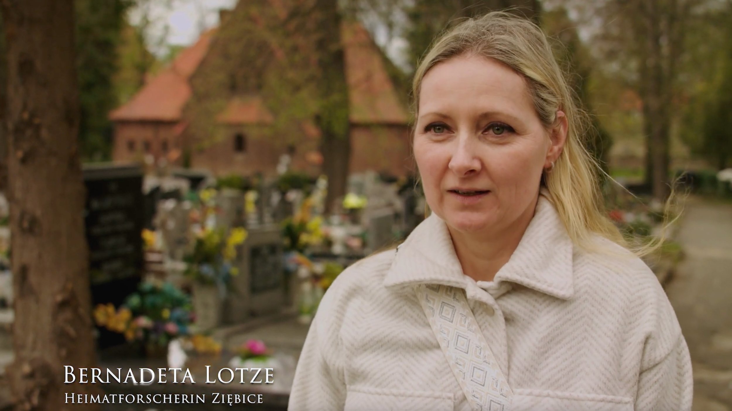
Dokumentar-Film "Karl Denke - Der Kannibale von Münsterberg (Ziębice)"

Dr. Mark Benecke als Experte für den Kannibalen-Fall Karl Denke 🦴

Karl Denke - der vergessene Kannibale - True Crime - History (2024)

Im Dezember 1924 scheitert Karl Denke am Versuch Vincenz Olivier mit einer Spitzhacke zu erschlagen. Er wäre sein 32. Opfer gewesen. Erstmalig rekonstruiert diese Dokumentation die Taten Karl Denkes an originalen Schauplätzen, mit unveröffentlichtem Bildmaterial, Filmausschnitten und kompetenten Interviewpartnern wie Dr. Mark Benecke und Armin Rütters.

47 Min., ab 16, Stream bei Amazon verfügbar


Strangest Things

UK & Deutschland | 2022


Serial killer Luis Alfredo Garavito Cubillos

Minerva Medicolegale | 2005


Serial killer Jürgen Bartsch

International Academy of Forensic Sciences (IAFS) | Montpellier, France


Der Macendoktor

Dokumentationsfilm


Gutachten: Überführen Maden

Bild am Sonntag | 1998

Halbes Bein im Volkspark gefunden

Quelle: Berliner Kurier, 1. März 2024, Seite 7

Von Stefanie Hildebrandt

Abgetrennter Oberschenkel: Kriminalbiologe Mark Benecke über den Gruselfund

In einem Park in Prenzlauer Berg wird ein Körperteil gefunden. Das verrät es dem Experten.

Die Suche nach weiteren Hinweisen im Fall eines abgetrennt aufgefundenen Oberschenkels in Berlin geht weiter und immer mehr Details werden bekannt. Bei der Berliner Polizei arbeitet man jedoch bisher im Verborgenen an dem Fall, hält sich mit Einzelheiten bedeckt.

Für den Berliner KURIER erklärt Deutschlands berühmtester Kriminalbiologe Mark Benecke, was sich für Experten alles an dem gefundenen Körperteil ablesen lässt.

In einem Park wird ein abgetrennter Oberschenkel gefunden. Was lässt sich an dem Fund ablesen?

“Anhand von Haut, Muskel, Haaren und dergleichen kannst du erst mal schauen, ob es eher ein Mann oder eher eine Frau sein könnte. Durch Erbgut kannst du das dann ebenfalls prüfen, aber auch schauen, ob die Person bekannt ist”, schreibt Mark Benecke. “Wenn sie beispielsweise schon mal in einer Erbgut-Datenbank eingestellt wurde, kriegst du einen “Treffer” und weißt, wer die Person ist von der der Schenkel stammt. Du kannst auch die Schnittkante anschauen: Kettensäge? Messer? Skalpell? Beil?”

Wie findet man heraus, wie lange der Schenkel schon da lag?

“Das geht nur über die Insekten-Besiedlung, das ist bei kaltem Wetter = wenigen Insekten aber schwierig. Zudem kann der Körperteil auch zwischendurch woanders gelegen haben, wo keine Insekten dran kommen, etwa einer Kühltruhe oder einer dicht verschlossenen Tüte oder Tasche.”

Wie kann man herausfinden, ob der Mensch lebte, als das Bein abgetrennt wurde?

„Durch Blutungen und Entzündungs-Merkmale an der Schnittkante. Wenn du lebst, blutet es im täglich bekannten Sinn‚ wenn jemand tot ist, dann verteilt sich das Blut kaum und anders. Im Gewebe-Dünn-Schnitt siehst du auch, ob zum Beginn den Versuch des Körpers zur Heilung gab, dann lebte die Person noch beim Schnitt. Wenn du keine Entzündungs- oder Heilungs-Merkmale in den Zellen siehst, war die Person schon tot”.

“Täter oder Täterinnen zerteilen Leichen öfter, um sie leichter tragen zu können, das heißt bei uns ‘defensive Leichen-Zerstückelung’, das heißt es hat nichts direkt mit der eigentlichen Tötung zu tun”, so Benecke.

Beim Abtrennen eines Oberschenkels werden große Blutgefäße durchtrennt, wie wahrscheinlich ist es, das bei laienhafter Ausführung zu überleben?

“Super unwahrscheinlich. Besonders die dicke Ader auf der Innenseite des Oberschenkels führt oft schon bei einem Stich hinein zum Verbluten.”

Einen Mordfall kann die Polizei in Berlin demnach auch nicht ausschließen.


Interview with Nico Claux

Cannibalism


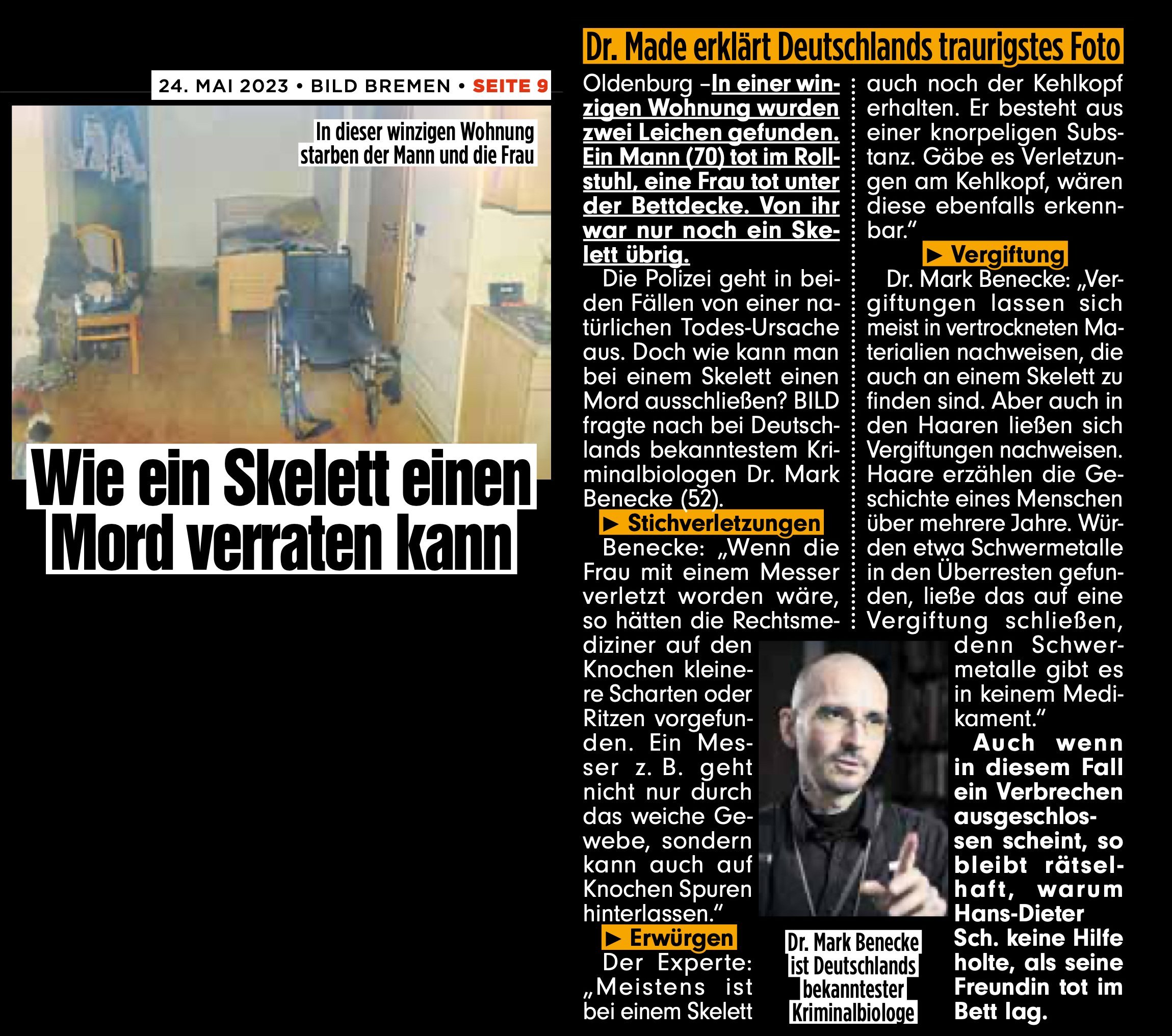
Wie ein Skelett einen Mord verraten kann

Bild 2023


Warum trank der Vampir von Düsseldorf Blut?

Bild 2023


Busen-Mord

Skurriler Kriminalfall aus Unna


Blutspuren, Serienmord, Forensik

Kurs an der Uni Siegen / Bonn


Zum Fall “Valeriia”

t-online | 2024

Über die Aussagekraft forensisch-entomologischer Untersuchungen bei nicht optimaler Asservierung und weiteren Defiziten

Zur Berechnung des Mindest-Besiedlungs-Zeitraumes von Fliegenmaden eines toten Hundes

Read More

Insects Under The Skin? Ziploc And Matchbox Evidence In The Expert Forensic Stain Laboratory

The idea of insects living inside and on the (human) body literally drives people affected by the thought crazy. We receive samples containing alleged insect stain evidence on a regular basis which on examination mostly turn out to be non-animal fragments of skin, hair, fibers et cetera.

Read More